Inverted Microscopes – IM-7 Series

The IM-7 Inverted Microscopes Series from Optika Microscopes stands at the forefront of research microscopy, offering unmatched innovation and expandability. Available from Apex Scientific in South Africa, this advanced platform is purpose-built for demanding life science applications.

Designed to meet all modern research requirements, the IM-7 is supported by specialized application packages and delivers state-of-the-art imaging performance. Its modular design ensures flexibility for future upgrades, making it a smart investment for evolving lab environments. Explore our range of laboratory solutions and visit our Insights page for expert guidance and resources.

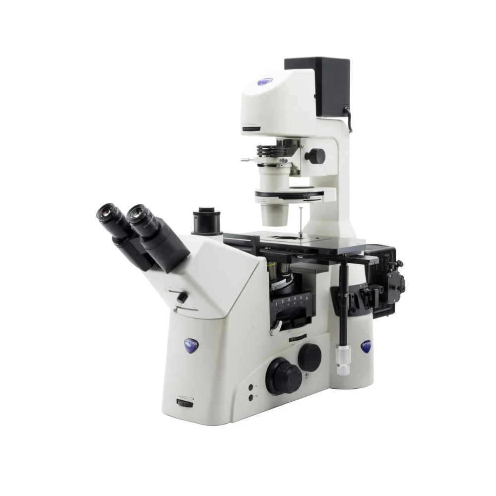
The IM-7 Inverted Microscopes Series from Optika Microscopes is a top-tier solution for advanced laboratories, available through Apex Scientific in South Africa. Specifically designed for life science research, this system combines innovation, modular expandability, and excellent imaging quality. Whether you’re performing detailed cellular observation or high-level manipulation, this inverted microscope offers the flexibility and performance you need.


Optical System and Head Design

The IM-7 Inverted Microscope features an infinity-corrected optical system that delivers consistently sharp, clear images. A trinocular head with a 45° inclination and a 47–78 mm interpupillary distance ensures ergonomic comfort. Furthermore, the built-in Bertrand lens improves image alignment, making this system ideal for complex research applications.


Versatile Photo Tubes and Eyepieces

This model provides flexible viewing options through Trino Port/Binocular settings of 100/0, 50/50, and 0/100. The PLAN WF10x eyepieces include a 25 mm high eyepoint and integrated rubber cups, offering user-friendly comfort during prolonged sessions.


Advanced Nosepiece and Objective Options

A sextuple ball-bearing revolving nosepiece with DIC slots in all positions supports a full range of objectives. Each lens is infinity-corrected, anti-fungus treated and offers a 60 mm parfocal distance. Users can select from multiple objective configurations to match their research goals.


Robust Specimen Stage

The three-layer mechanical stage, sized at 340×230 mm with a 130×85 mm movement range, uses a rack-and-pinion mechanism for precise navigation. It includes holders for Petri dishes and Terasaki plates, accommodating various experimental formats with ease.


Precision Focusing and Reliable Illumination

The IM-7 Inverted Microscope includes coaxial coarse and fine focus with tension control and limit stops to protect your samples. Its Koehler-type condenser offers phase contrast and a 26 mm working distance. With X-LED10 illumination, you benefit from high light output, energy efficiency, and a 65,000-hour service life.


Modular Upgrades for Advanced Imaging

For even more capabilities, the IM-7 can be upgraded with incident fluorescence and Differential Interference Contrast (DIC). These options transform the system into a versatile imaging platform suitable for both basic and advanced research techniques.

To help you select the best configuration, read our article, Choosing the Right Microscope for Your Laboratory: A Detailed Guide. You’ll also find helpful tips and educational content on our Insights page, or you can browse our complete product range for other laboratory equipment available in South Africa.

TechniqueDescriptionExample
BrightfieldTransmitted brightfield illumination is one of the most commonly used observation methods in optical microscopy, and is ideal for fixed, stained specimens or other types of samples having high natural absorption of visible light.
IM-7 is fitted with high-efficiency LED brightfield illuminator, for the best outcome when using this technique.
Capsella middle embry – Brightfield
DICDifferential Interference Contrast (DIC) is a microscopy technique that introduces contrast to images of specimens which have little or no contrast when observed using brightfield microscopy. The images produced using DIC have a pseudo 3D-effect, making the technique ideal for many applications.
DIC produces high resolution images with good contrast. It is best for observing unstained samples.
Sphagnum pores g – DIC
FluorescenceThe fluorescence microscopy is the most demanding technique in biology and biomedical sciences, as well as in materials science.
This method is capable to study organic and inorganic samples thanks to primary fluorescence (auto-fluorescence) or secondary (staining and labelling with fluorochromes).
IM-7 is tailored for applications in research, clinical and pharmaceutical diagnostic field.
Fluorescence illuminators available as mercury lamp.
Cotton fibers – UV Fluorescence
Phase ContrastPhase-contrast microscopy is a particular technique applied in transparent, non-stainable samples like culture of living cells, microorganisms, lithographic patterns, latex dispersions, fibers, asbestos, and subcellular particles.
It reveals many cellular structures that are not visible with a simple brightfield microscope.
Diatoms – Phase contrast


IM-7 – Objectives/Application Packages

Objectives

CodeDescription
M-1320IOS U-PLAN F (SEMI-APO) PH 4x/0.13, W.D. 16.5 mm, Cover glass
M-1321IOS U-PLAN F (SEMI-APO) PH 10x/0.3, W.D. 7.4 mm, Cover glass 1.2 mm
M-1322IOS U-PLAN F (SEMI-APO) PH 20x/0.45, W.D. 7.5–8.8 mm, Cover glass 0–2 mm
M-1323IOS U-PLAN F (SEMI-APO) PH 40x/0.60, W.D. 3.0–4.4 mm, Cover glass 0–2 mm
M-1324IOS U-PLAN F (SEMI-APO) PH 60x/0.70, W.D. 1.8–2.6 mm, Cover glass 0.1–1.3 mm
M-1358IOS U PLAN APO Objective 60x/1.40 (Oil), W.D. 0.25 mm, Cover glass 0.17 mm


HBO Fluorescence Package

CodeDescription
M-1330EPI Fluorescence internal attachment
M-151.1OSRAM 100W HBO high pressure mercury bulb
M-1332HBO Lamp house
PS-HBOOptika 100W HBO power supply
M-13346-position fluorescence filterbox turret
M-1335UV protector orange shield
M-1336B filterblock, filters included
M-1337G filterblock, filters included
M-1338V filterblock, filters included
M-1339UV filterblock, filters included
M-1340Aperture diaphragm slider
M-1341Field diaphragm slider
M-1342Slider with neutral density filter for HBO illumination
M-1343Empty fluorescence filterblock


Fluorescence Filter Specifications

CodeColorExcitation Filter (nm)Dichroic Cut-off (nm)Emission Filter (nm)
M-1336B (Blue)460 – 490505515LP
M-1337G (Green)510 – 550570575LP
M-1338V (Violet)385 – 425440455LP
M-1339UV (Ultraviolet)325 – 375415435LP


DIC – Differential Interference Contrast Package

CodeDescription
M-1350DIC Prism for 10x
M-1351DIC Prism for 20x
M-1352DIC Prism for 40x
M-1353DIC Prism for 60x
M-1354DIC 1 prism 10x for condenser
M-1355DIC 2 prism 20x-40x-60x for condenser
M-1356Slider with rotating analyzer


PH – Phase Contrast Package

CodeDescription
M-13484X Phase ring
M-134960X Phase ring


Product Enquiry

"*" indicates required fields

First Name*
Last Name*