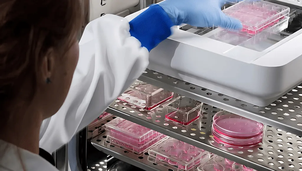
Live cell imaging has transformed the way scientists monitor and study cells in real-time. Whether you are researching cell proliferation, conducting scratch assays for wound healing, or observing organoids, understanding the technical specifications and capabilities of live cell imaging systems is essential for selecting the right tool for your research. This article explores key factors to consider when choosing a live cell imaging system and how devices like Axion Biosystems’ Omni outperform others in the market.
What is Live Cell Imaging?
Live cell imaging refers to the use of advanced imaging technology to observe the behavior and interactions of living cells in real time. This allows researchers to track dynamic cellular processes such as cell division, migration, and apoptosis under physiological conditions. With real-time insights, live cell imaging is invaluable for studying the biological mechanisms of cell proliferation, wound healing, and more.
Key Applications
The ability to observe live cells has opened up new avenues in research. Some of the most common applications include:
- Cell Proliferation Studies: Monitoring cell growth and division is crucial in cancer research, stem cell therapy, and drug development.
- Scratch Assay: This technique is used to study cell migration and wound healing, important for areas like tissue engineering and regenerative medicine.
- Organoids: Live cell imaging is vital for studying 3D cell culture systems, like organoids, which replicate the structure and function of real tissues. These are increasingly used for drug screening, disease modeling, and regenerative research.
As a laboratory equipment supplier in South Africa, Apex Scientific provides a wide range of live cell imaging systems, including solutions for cell proliferation studies and organoid imaging.
Why Choose Live Cell Imaging Over a Microscope?
While traditional microscopes have long been used for cell observation, they have limitations when it comes to monitoring dynamic cellular processes. Here’s why these systems are superior:
- Real-time Monitoring: Unlike conventional microscopes, live cell imaging systems allow continuous observation, enabling you to track cellular events over time.
- High Throughput: With live cell imaging, multiple samples can be observed simultaneously, ideal for large-scale experiments and high-content screening.
- Non-invasive: These systems offer non-invasive observation, reducing the need for cell labeling or treatments that may affect cell health during experiments.
What Sets the Omni Apart from Other Devices?
- Superior Imaging Resolution: The Omni provides high-resolution imaging, ensuring that even the smallest cellular details are captured with clarity.
- Multifunctionality: It is suitable for various applications, such as cell proliferation, scratch assays, and organoid imaging, making it a versatile tool for any laboratory.
- Cutting-edge Technology: Equipped with the latest advancements, the Omni offers precise real-time imaging, giving researchers the ability to track live cellular processes with accuracy.
- User-friendly Interface: The Omni’s intuitive interface makes it accessible to both novice and experienced researchers, simplifying the imaging process.
- Long-term Cell Culture Compatibility: The Omni is optimized for long-term experiments, providing controlled environments that keep cells healthy during extended imaging sessions.
Important Technical Specifications to Consider
Selecting the right system requires careful attention to various specifications:
- Imaging Resolution: Higher resolution ensures better clarity of cellular details, which is crucial for studying fine cellular structures.
- Frame Rate: A high frame rate is necessary to capture fast cellular events like cell division and migration.
- Multichannel Imaging: Systems with multichannel capabilities allow researchers to monitor different cellular processes simultaneously.
- Temperature and Environmental Control: Maintaining optimal conditions for live cells is critical, especially for long-term imaging.
- Software Features: Software that supports automated image analysis, real-time monitoring, and high-content screening is essential for efficient data processing.
Choosing the Right Accessories
Many systems come with accessories that enhance their functionality. Some key accessories include:
- Custom Fluorescence Filters: Specific applications, such as tracking proteins or other cellular components, require the right filters for optimal results.
- Environmental Chambers: These are essential for maintaining proper growth conditions for cells, especially during extended imaging periods.
- Sample Holders and Stages: Choosing the right sample holder ensures stable and accurate positioning of samples for optimal imaging.
Conclusion
Choosing the right live cell imaging system is essential for obtaining reliable, high-quality results in your research. By understanding the key specifications and applications, such as cell proliferation and organoid studies, you can make an informed decision. With systems like the Omni, researchers gain the ability to track dynamic cellular processes with unmatched precision, ensuring successful outcomes in experiments related to wound healing, scratch assays, and beyond.
For more information about live cell imaging systems available in South Africa, visit Apex Scientific.


